Consumer panels and sauna-plus-red-light rooms outran the dose literature: the mechanism and the limit
Clinical photobiomodulation requires specific wavelengths (typically 630–670 nm and 810–850 nm) delivered at a controlled energy density in joules per square centimeter; sitting near an uncalibrated consumer panel or sauna light fixture rarely reproduces a clinical trial dose.
How wavelength, skin-level irradiance, and tissue penetration separate medical LED guides from gym panels and sauna rooms.
LetsGo Care Editorial
Consumer panels and sauna-plus-red-light rooms outran the dose literature: the mechanism and the limit
Sitting near a glowing red panel in a recovery studio or sauna does not automatically reproduce the photon dose used in clinical photobiomodulation trials.
Red light therapy, known in medical literature as photobiomodulation (PBM) or low-level light therapy (LLLT), uses non-ionizing light in the visible red (620–700 nm) and near-infrared (780–1100 nm) spectrums to alter cellular function without heating tissue. While dermatology clinics and physical therapy researchers use calibrated light-emitting diodes (LEDs) or low-power lasers at exact distances and exposure times, commercial wellness facilities increasingly bundle uncalibrated red light panels into saunas and recovery rooms. Understanding the physics of light delivery—specifically irradiance at the skin surface and the biphasic dose response—explains why targeted clinical guides show measurable benefits for skin and joint tissue while ambient full-body exposure often yields no systemic change.
Key facts
- Primary mechanism: Photons between 630–670 nm and 810–850 nm are absorbed by cytochrome c oxidase (Complex IV) in the mitochondrial electron transport chain, displacing inhibitory nitric oxide and transiently increasing ATP synthesis in cell and animal models.
- Biphasic dose response (Arndt-Schulz curve): Too little energy density (fluence, measured in J/cm2) produces no biological effect, an optimal window stimulates tissue repair, and excessive fluence inhibits cellular function or causes thermal stress.
- Clinical evidence is strongest for localized dermatological applications (androgenetic alopecia, photoaged skin) and superficial musculoskeletal pain (knee osteoarthritis, tendinopathy) in randomized controlled trials of 4 to 24 weeks.
- Distance and reflection dictate dose: Light from non-collimated LED panels obeys the inverse-square law, and roughly 40% to 60% of incident photons reflect off the stratum corneum unless the emitter is placed close to or in direct contact with bare skin.
- Systemic claims lack human data: In-human randomized trials have not established that full-body red light panels lower systemic inflammatory markers like hs-CRP, alter epigenetic BioAge clocks, or improve resting metabolic rate.
Who this is for
Adults aged 40 to 65 managing localized osteoarthritis pain, tendinopathy, androgenetic alopecia, or facial photoaging who are willing to apply a calibrated device at a fixed distance and schedule, rather than those seeking passive systemic metabolic or longevity effects from ambient sauna lighting.
In this article
- What it is
- Why it is used
- How it helps
- Who should try it
- Measurements that show improvement
- Products and services
What it is
Photobiomodulation (PBM) is the non-thermal application of monochromatic or narrow-band light to biological tissues. Unlike intense pulsed light (IPL) or ablative dermatological lasers, which rely on selective photothermolysis to destroy targets by heating water, hemoglobin, or melanin, true PBM operates at power densities that do not raise tissue temperature by more than 1°C.
Most evidence-based guides use two specific optical windows where absorption by water and hemoglobin is relatively low, allowing photons to penetrate past the epidermis:
- Visible red light (primarily 630 nm to 670 nm): Penetrates roughly 1 to 3 millimeters into the skin, absorbed heavily in the epidermis and dermis.
- Near-infrared (NIR) light (primarily 810 nm to 850 nm): Penetrates roughly 3 to 10 millimeters in human tissue, reaching subcutaneous fat, superficial tendon sheaths, and small joint capsules. Claims of deep organ or deep brain penetration in humans from external consumer LED panels are physically implausible; cadaver and in vivo human dosimetry studies demonstrate that over 90% of 810 nm light is scattered or absorbed within the first centimeter of tissue.
A clinical PBM prescription is defined by two parameters that consumer rooms rarely disclose:
- Irradiance (power density): Measured in milliwatts per square centimeter (mW/cm2) at the actual surface of the skin, not at the surface of the bulb. Consumer panel manufacturers frequently measure irradiance with a solar power meter (which overestimates narrow-band LED output by 50% to 100% compared to a calibrated optical spectrometer) and take the reading at zero distance.
- Fluence (energy density or dose): Measured in joules per square centimeter (J/cm2), calculated as Irradiance (W/cm2) multiplied by Time (seconds). Superficial skin targets typically respond to 2 to 10 J/cm2 at the skin surface, whereas deeper joint or tendon targets require 20 to 60 J/cm2 at the skin surface so that 1 to 5 J/cm2 actually reaches the target depth.
When a red light panel is mounted inside an infrared or traditional sauna, two physical problems occur. First, standing or sitting 60 to 100 centimeters away from a non-collimated LED array drops the irradiance at the skin to a fraction of its rated output, often requiring 30 to 60 minutes just to reach a minimal 4 J/cm2 threshold. Second, sauna heat induces immediate cutaneous vasodilation and sweating; increased dermal blood volume (hemoglobin) and a layer of sweat absorb and scatter incoming red photons in the superficial skin, preventing deeper penetration and confounding the non-thermal mitochondrial mechanism with massive thermal stress.
Why it is used
In clinical and high-agency self-care settings, red and near-infrared light are used across four distinct categories, ordered here from strongest to weakest human evidence:
-
Dermatology and Trichology (Established Human Evidence): Used to increase dermal collagen density, reduce fine periorbital rhytids (wrinkles), calm inflammatory acne vulgaris (often combined with blue 415 nm light), and stimulate hair follicle cycling in androgenetic alopecia (male and female pattern hair loss). Several low-level laser and LED helmets/combs hold FDA 510(k) clearance for androgenetic alopecia.
-
Supportive Oncology Care (Established Clinical Guideline): Intraoral red light (630–660 nm) and extraoral NIR light are recommended in clinical practice guidelines by the Multinational Association of Supportive Care in Cancer (MASCC) and the International Society of Oral Oncology (ISOO) to prevent and treat oral mucositis in patients undergoing hematopoietic stem cell transplantation or head and neck radiotherapy.
-
Superficial Musculoskeletal Pain and Exercise Recovery (Moderate Human Evidence): Applied directly over the knee joint line, Achilles tendon, lateral epicondyle, or cervical spine to reduce pain scores in osteoarthritis and tendinopathy. In sports physiology, pre-exercise application to specific muscle groups (such as the quadriceps) is used to delay local muscular fatigue and modestly attenuate post-exercise creatine kinase (CK) spikes.
-
Sleep, Mood, and Systemic Longevity (Emerging or Weak Human Evidence): Consumer marketing heavily promotes full-body red light for circadian rhythm entrainment, testosterone elevation, systemic inflammation reduction, and systemic longevity. Aside from small pilot trials suggesting that evening ambient red light avoids the melatonin suppression caused by blue/green room lighting, clinical trials have not demonstrated that skin exposure to red light increases human serum testosterone or extends healthspan.
How it helps
The primary photoacceptor for red and near-infrared light in mammalian cells is cytochrome c oxidase (CCO), which is Complex IV of the mitochondrial respiratory chain. This mechanism is well established in isolated mitochondria, cell culture, and rodent models, with indirect confirmation in human tissue via near-infrared spectroscopy (NIRS).
-
Cytochrome c Oxidase and Nitric Oxide Photodissociation: CCO contains two heme centers (heme a and heme a3) and two copper centers (CuA and CuB) that absorb photons in the 620–680 nm and 760–895 nm bands. Under conditions of cellular stress, hypoxia, or inflammation, nitric oxide (NO) binds competitively to the reduced binuclear center (heme a3/CuB) of CCO, competitively inhibiting oxygen binding and slowing electron transport. Absorption of red or NIR photons breaks the photodissociable bond between NO and the metal centers. Once NO is displaced, oxygen binds freely, electron transport resumes, the proton gradient across the inner mitochondrial membrane increases, and adenosine triphosphate (ATP) synthesis rises.
-
Transient Reactive Oxygen Species (ROS) and Transcription Factor Signalling: The brief burst in mitochondrial electron transport generates a small, transient pulse of reactive oxygen species (ROS). In healthy cells, this low-level ROS burst acts as a retrograde signal from the mitochondria to the nucleus, activating redox-sensitive transcription factors including nuclear factor kappa B (NF-kB) and nuclear factor erythroid 2-related factor 2 (Nrf2). This triggers the upregulation of endogenous antioxidant enzymes (such as superoxide dismutase and glutathione peroxidase) and cytoprotective genes. Simultaneously, the displaced free nitric oxide acts as a local vasodilator via cyclic GMP (cGMP), improving microcirculation in the irradiated tissue.
-
Light-Gated Ion Channels and TGF-beta1: A secondary mechanism identified in cell studies involves light-sensitive transient receptor potential (TRP) channels, particularly TRPV1, primarily at longer wavelengths (>900 nm) where intracellular water clusters absorb photon energy. In gingival and dermal tissues, PBM also activates latent transforming growth factor-beta 1 (TGF-beta1), driving fibroblast proliferation and pro-collagen synthesis while downregulating matrix metalloproteinases (MMP-1 and MMP-2) that degrade extracellular matrix.
Non-Obvious Synthesis—Why Local Markers Move While Systemic Outcomes Do Not: Photobiomodulation strictly obeys the Arndt-Schulz biphasic dose response curve. If the photon dose is too low, CCO inhibition is not overcome. If the dose is within the therapeutic window, ATP and protective signalling rise. If the dose is too high, excessive ROS production overwhelms endogenous antioxidants, opening the mitochondrial permeability transition pore and inhibiting cellular activity.
Because human skin and subcutaneous tissue scatter photons exponentially, it is physically impossible for a single flat panel to deliver an optimal Arndt-Schulz dose to the epidermis, the underlying skeletal muscle, and deeper vasculature simultaneously. A dose high enough to deliver 3 J/cm2 to the vastus lateralis muscle belly at 2 centimeters depth will deliver 50 to 100 J/cm2 to the overlying epidermis—pushing the skin into the inhibitory zone of the biphasic curve. Conversely, a panel placed 50 centimeters away delivers a mild, non-inhibitory dose to the skin (3–5 J/cm2) that extinguishes completely before reaching skeletal muscle or major blood vessels. Therefore, biopsy and localized imaging markers in human trials consistently show dermal collagen remodeling or superficial joint analgesia when dosed appropriately for depth, while systemic blood markers (such as systemic hs-CRP, fasting insulin, or leukocyte epigenetic clocks) remain flat. The photon energy simply does not reach enough total body cell mass at an effective fluence to drive a systemic metabolic shift.
Who should try it
Human clinical trial data support targeted red and near-infrared light therapy for specific, localized presentations rather than general wellness:
- Androgenetic Alopecia: Systematic reviews and meta-analyses of randomized, sham-controlled trials (typically 16 to 26 weeks in duration, enrolling 40 to 250 adults per trial using 650–660 nm laser/LED helmets or combs 3 to 7 times per week) show a statistically significant increase in terminal hair density of roughly 15 to 20 hairs/cm2 compared to sham devices. Effect sizes are comparable to topical minoxidil 2%–5%, and combination therapy outperforms monotherapy.
- Facial Photoaging and Dermal Density: Split-face randomized controlled trials (typically 30 to 120 participants over 8 to 12 weeks, using 630–660 nm alone or combined with 830 nm at 5 to 20 J/cm2 twice weekly) demonstrate measurable reductions in periorbital wrinkle depth on 3D optical profilometry and increased intradermal collagen density on high-resolution ultrasound.
- Knee Osteoarthritis and Superficial Tendinopathy: Meta-analyses of randomized placebo-controlled trials (covering over 1,000 patients with knee osteoarthritis) indicate that 810–850 nm or 904 nm light lowers Visual Analog Scale (VAS) pain scores by roughly 12 to 20 mm (on a 100 mm scale) and improves WOMAC disability scores over 4 to 8 weeks—but only when the device is applied in direct skin contact over the joint capsule at doses adhering to World Association for Photobiomodulation Therapy (WALT) guidelines (typically 4 to 8 J per point at 810 nm). Trials using non-contact panels or under-dosed probes show no benefit over sham.
- Local Muscular Recovery: Randomized crossover trials in trained and untrained adults show that applying 660/850 nm clusters in direct contact with the quadriceps or biceps immediately before resistance exercise increases repetitions to failure by roughly 5% to 10% and blunts 24–48 hour delayed-onset muscle soreness (DOMS).
Trade-offs, Contraindications, and Opportunity Cost:
- Side effects: Transient erythema (skin redness), mild dryness, temporary ocular afterimages or headache if eye protection is omitted, and paradox inhibition (increased local fatigue or stalled hair growth) if over-dosed daily.
- Pigment interactions: Melanin is a potent absorber of visible red light (630–670 nm). Individuals with darker skin phototypes (Fitzpatrick IV–VI) absorb more energy in the basal epidermis, which reduces deeper penetration and carries a documented risk of post-inflammatory hyperpigmentation or melasma flare if high-irradiance visible red light generates mild epidermal heating. Near-infrared (810–850 nm) is less absorbed by melanin, though caution in active melasma is still advised.
- Drug interactions: Concurrent use of photosensitizing medications—including tetracyclines (doxycycline), fluoroquinolones, isotretinoin, amiodarone, hydrochlorothiazide, phenothiazines, or topical retinoids applied immediately prior to exposure—can trigger phototoxic skin reactions.
- Contraindications: Never apply directly over known or suspected cutaneous malignancies (basal cell carcinoma, squamous cell carcinoma, melanoma) or active systemic tumors, as in vitro data show PBM can stimulate proliferation in certain dysplastic cell lines. Avoid direct application over the thyroid gland in individuals with hyperthyroidism or autoimmune thyroiditis unless supervised in a clinical trial, and avoid over the gravid uterus during pregnancy.
- Cost and opportunity cost: Calibrated home panels and medical-grade hair helmets cost $400 to $3,000; studio memberships run $100 to $300 monthly. Spending 20 minutes daily standing in front of a panel carries a direct time opportunity cost if it displaces zone 2 aerobic conditioning, progressive resistance training, or sleep duration—interventions that produce established, large-magnitude improvements in VO2max, insulin sensitivity, and all-cause mortality.
Measurements that show improvement
Because photobiomodulation acts locally, tracking systemic longevity blood panels (such as ApoB, HbA1c, fasting insulin, or systemic hs-CRP) will not reflect whether a red light guide is working. If a reader integrates red light therapy into a structured LetsGo tracking program, the measurable markers must match the targeted tissue:
- Hair Density and Shaft Diameter (for Scalp guides):
- Measurement: Standardized macro-photography or trichoscopy (phototrichogram) at a fixed anatomical landmark (such as the vertex or frontal hairline intersection) at starting point, 16 weeks, and 26 weeks.
- Direction of change: Stabilization of starting point hair count (halting progressive miniaturization) or an increase in terminal hair density (hairs/cm2) and mean hair shaft thickness.
- Standardized Pain and Joint Function Scores (for Osteoarthritis or Tendinopathy):
- Measurement: 0–100 mm Visual Analog Scale (VAS) for pain during a standardized load (such as a single-leg decline squat for patellar tendinopathy or stair descent for knee OA) and the Western Ontario and McMaster Universities Osteoarthritis Index (WOMAC), tracked weekly over 4 to 8 weeks.
- Direction of change: A reduction of at least 15–20 mm on the VAS (the minimal clinically important difference) and increased pain-free range of motion.
- Dermal Elasticity and Pigmentation Uniformity (for Facial guides):
- Measurement: Standardized cross-polarized clinical photography (such as VISIA skin analysis) or cutometer elasticity readings taken at starting point and 12 weeks.
- Direction of change: Reduction in fine rhytid count and periorbital erythema; an increase in brown spot counts indicates melanin activation/hyperpigmentation and signals that the guide should be halted.
- Local Neuromuscular Recovery and Sleep Architecture:
- Measurement: Force plate countermovement jump height or isometric mid-thigh pull recovery 24 hours after eccentric lower-body training; overnight heart rate variability (HRV, specifically RMSSD) and sleep onset latency via a validated wearable if evening ambient red light is used to replace overhead white/blue lighting.
- Direction of change: Faster return to starting point peak torque within 24–48 hours post-exercise; shortened sleep onset latency (by 5 to 15 minutes) when evening melanopic lux exposure is reduced.
Before tracking biological outcomes, verify the physical dose input: confirm the device emits narrow-band peaks around 630–670 nm and/or 810–850 nm, find its true spectrometer-tested irradiance (typically 20 to 60 mW/cm2 at 15 cm for quality LED panels), and calculate exposure time so each target area receives 4 to 10 J/cm2 for skin or 20 to 40 J/cm2 for superficial joints, delivered 3 to 5 times per week rather than multiple times per day.
Products and services
These LetsGo products and services sit next to this subject. They are not a prescription. A link is not evidence that the product causes the change discussed above.
-
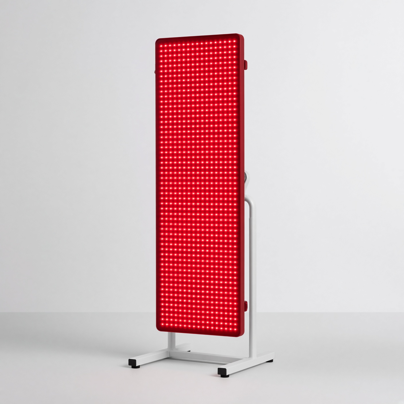
Red light therapy panel — A panel for clinics and home recovery. Distance and time matter more than the brochure.
-
LetsGo programmes — Coached programmes. Not a clinic menu and not a promise of a result.
Frequently asked questions
Does combining red light therapy with a sauna improve results?
No clinical evidence shows synergy between sauna heat and photobiomodulation, and the physics of light transport suggest they conflict. Photobiomodulation relies on non-thermal photon absorption by cytochrome c oxidase. Sauna heat causes intense skin vasodilation (increasing hemoglobin, a competing chromophore that absorbs red light) and coats the skin in sweat, which reflects and scatters photons. Furthermore, panels mounted on sauna walls are usually too far from the body to deliver a therapeutic fluence (J/cm2), and extreme ambient heat degrades LED diode efficiency and lifespan.
Can red light therapy penetrate clothes or sunscreen?
No. Clothing fibers, mineral sunscreens (zinc oxide and titanium dioxide), and cosmetic foundations reflect, scatter, and absorb both visible red and near-infrared light. For clinical dosing, the light source must be applied to clean, bare skin with no topical barriers.
Are LED panels as effective as clinical lasers for photobiomodulation?
For superficial tissues like the epidermis, dermis, and scalp hair follicles, narrow-band LEDs (630–660 nm and 810–850 nm) produce biological outcomes comparable to low-level lasers when matched for wavelength, irradiance, and total fluence (J/cm2), because laser coherence is lost immediately upon scattering in the stratum corneum. However, for deeper tendons and joint capsules (such as knee osteoarthritis), clinical lasers or cluster probes pressed directly into the skin with firm contact displace superficial blood and achieve better photon penetration than non-contact LED panels held several inches away.
How long should a red light therapy session last?
Session duration depends entirely on the true irradiance (mW/cm2) at the skin surface and the target tissue depth. At a verified skin irradiance of 30 mW/cm2 (0.03 W/cm2), delivering a superficial skin dose of 6 J/cm2 takes 200 seconds (just over 3 minutes). Delivering a deeper musculoskeletal surface dose of 30 J/cm2 at that same irradiance takes 1,000 seconds (about 16.5 minutes). Exceeding 15 to 20 minutes per area at close range frequently pushes tissues into the inhibitory phase of the biphasic dose response, reducing cellular ATP output.
Practical takeaways
- Separate heat from light: Use saunas for cardiovascular and heat-shock protein conditioning, and perform photobiomodulation separately on cool, dry, bare skin so sweat and cutaneous vasodilation do not block photon penetration.
- Calculate your dose in J/cm2: Demand spectrometer-tested irradiance numbers (not solar meter readings) from the device manufacturer. Multiply irradiance in W/cm2 by exposure time in seconds to hit 4–10 J/cm2 for skin/scalp or 20–40 J/cm2 for superficial joints.
- Control distance and contact: For joint and tendon pain, use a device designed for direct skin contact or hold the emitter within 2 to 5 centimeters of the joint line; for facial skin or scalp, maintain a consistent distance (typically 10 to 20 cm for panels, or direct fit for helmets) across every session.
- Respect the biphasic curve: Limit exposure to 3 to 5 sessions per week per anatomical site. Daily over-exposure reduces clinical efficacy via excess reactive oxygen species.
- Protect the eyes and pigmented skin: Wear opaque goggles when treating the face with high-output LEDs, especially when near-infrared (810–850 nm) wavelengths are active (as NIR does not trigger the blink reflex), and avoid visible red light on areas prone to melasma.
Sources and evidence notes
- Hamblin MR. Mechanisms and Mitochondrial Redox Signaling in Photobiomodulation (Photochemistry and Photobiology, 2018). Details cytochrome c oxidase photon absorption, nitric oxide photodissociation, retrograde ROS signalling, and light-gated ion channel mechanisms in cellular and animal models.
- Huang YY, Sharma SK, Carroll J, Hamblin MR. Biphasic Dose Response in Low Level Light Therapy - An Update (Dose-Response, 2011). Establishes the Arndt-Schulz biphasic fluence curve demonstrating why excessive irradiance or exposure time inhibits ATP synthesis and tissue repair.
- Zadik Y, Arany PR, Fregnani ER, et al. Systematic review of photobiomodulation for the management of oral mucositis in cancer patients and clinical practice guidelines (Supportive Care in Cancer, MASCC/ISOO, 2019). Provides high-grade human RCT evidence and exact wavelength/fluence dosing guidelines for mucosal tissue protection.
- Stausholm MB, Naterstad IF, Joensen J, et al. Efficacy of low-level laser therapy on pain and disability in knee osteoarthritis: systematic review and meta-analysis of randomised placebo-controlled trials (BMJ Open, 2019). Meta-analysis of 22 RCTs (1,063 patients) demonstrating that pain reduction in human knee osteoarthritis occurs only when wavelengths and doses match WALT clinical thresholds.
- Adil A, Godwin M. The effectiveness of treatments for androgenetic alopecia: A systematic review and meta-analysis (Journal of the American Academy of Dermatology, 2017). Quantifies human RCT effect sizes for low-level light therapy in male and female pattern hair loss compared against minoxidil and finasteride.
Safety note
Do not apply red or near-infrared light over active skin cancers, suspicious pigmented lesions, or the thyroid gland. Discontinue use if you take photosensitizing medications or develop hyperpigmentation. Always wear wavelength-appropriate eye protection when emitters are directed near the face, as near-infrared light (810–850 nm) is invisible to the human eye and does not trigger a protective blink or pupillary constriction reflex.
Disclaimer
This article is for educational purposes only and does not constitute medical advice, diagnosis, or a treatment prescription. Photobiomodulation parameters vary by clinical condition and individual skin phototype; consult a qualified physician or dermatologist before starting light therapy for a medical or dermatological condition.
Red and near-infrared light therapy is a biochemically grounded, localized intervention whose clinical outcomes depend entirely on optical physics. When wavelengths of 630–670 nm or 810–850 nm are delivered to bare skin at verified irradiances and appropriate fluences, human randomized trials confirm meaningful improvements in androgenetic alopecia, dermal collagen architecture, oral mucositis, and superficial joint or tendon pain. Conversely, passive exposure to uncalibrated red light panels in saunas or recovery rooms violates the physical requirements of photon penetration and the biphasic dose response. High-agency adults get clinical value from photobiomodulation by treating it as a targeted, dose-specific tissue therapy rather than a systemic longevity shortcut.